top of page
Outwardly propagating Ca wave propagation is not blocked by localized BAPTA/AM Ca buffering
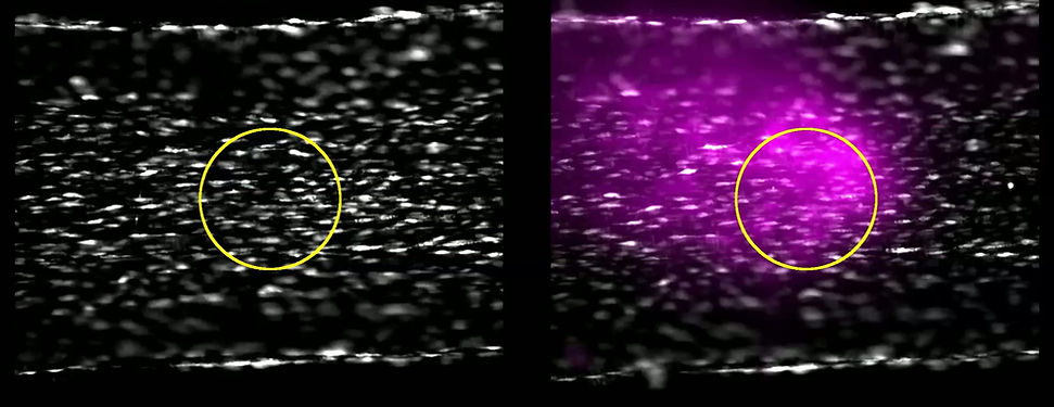
En face mesenteric artery stained with Cal520/AM (5 µM, grey) and cIP (5 μM). Ca activity was stimulated by localized photolysis of cIP (yellow circle) after a baseline recording (5 Hz per channel; baseline cut short for the purpose of video). In the same preparation, the resulting Ca activity is visualized in green before (control) and after localized BAPTA/AM (30 µM, 15 mins plus 15 mins rest) application using pressure ejection from a puffer pipette, determined using sulforhodamine B (1 µM) signal (magenta application region). Imaging was performed in the presence of counter-propagating PSS flow (1.5 ml.min-1). Scale bar: 50 μm.
2+
2+
2+
2+
3
3
bottom of page