Smooth Muscle Cells

Smooth Muscle


Isolated smooth muscle cells
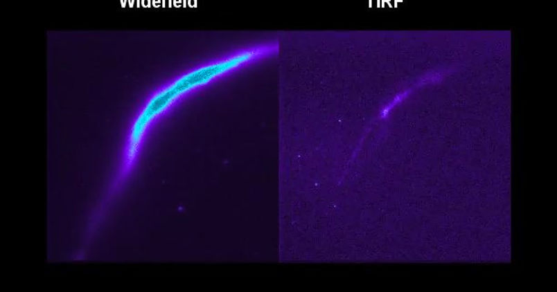
Smooth muscle cells allow arteries to rapidly contract and relax to alter blood flow, ultimately contributing to regulation of blood pressure. To dynamically alter luminal diameter, smooth muscle cells respond to IP3-generating agonists by producing Ca2+ waves, visualised in the video above. In this instance, carbachol was used to initiate a uniform rise in cytoplasmic Ca2+ concentration over a single, though substantial, length of the cell (~30um). In order to finely control experimental conditions, we have the ability to patch the primary smooth muscle cell, while bringing a patch pipette close with which to administer drugs or other reagents.
Atherosclerotic plaques are populated with smooth muscle cells (SMCs) and macrophages. SMCs are thought to accumulate in plaques because fully differentiated, contractile SMCs reprogramme into a ‘synthetic’ migratory phenotype, so-called phenotypic modulation, whilst plaque macrophages are thought to derive from blood-borne myeloid cells. Following the fate of SMCs is complicated by the lack of specific markers for the migratory phenotype, therefore we employed long-term, high-resolution, time-lapse microscopy to track the fate of unambiguously identified, fully-differentiated, contractile SMCs in response to the growth factors present in serum.

SMC transform and migrate
Phenotypic modulation can be clearly seen in this brightfield timelapse video. The highly elongated, contractile SMCs initially rounded up before spreading outwards. Once spread, the SMCs became motile and displayed dynamic cell-cell communication behaviours, displaying clear evidence of phagocytic activity. Resident SMCs may provide a potential source of macrophages in vascular remodelling.
Flicker-assisted localization microscopy, FaLM, provides a convenient tool for determining mitochondrial architecture and has revealed smooth muscle cell mitochondrial alterations that may contribute to hypertension.
Mitochondrial morphology is central to normal physiology and disease development. However, in many live cells and tissues, complex mitochondrial structures exist and morphology has been difficult to quantify.

We have measured the shape of electrically-discrete mitochondria, imaging them individually to restore detail hidden in clusters and demarcate functional boundaries. Stochastic “flickers” of mitochondrial membrane potential were visualized with a rapidly-partitioning fluorophore and the pixel-by-pixel covariance of spatio-temporal fluorescence changes analyzed. This Flicker-assisted Localization Microscopy (FaLM) requires only an epifluorescence microscope and sensitive camera. In vascular myocytes, the apparent variation in mitochondrial size was partly explained by densely-packed small mitochondria. In normotensive animals, mitochondria were small spheres or rods. In hypertension, mitochondria were larger, occupied more of the cell volume and were more densely clustered.

Intact VSMCs
We can also visualise VSMC activity in intact mesenteric artery preparations. In this video, VSMCs have been loaded up with the Ca2+ indicator dye Cal520 and are visualised using an EMCCD camera on an upright inverted microscope.
Voltage dependent calcium channels (VDCCs) are a group of membrane bound ion channels with permeability to ion Ca2+ and activated at depolarized membrane potential.
Calcium influx is triggered by activating VDCCs with perfusing high K+ (20 mM) PSS into the bath of preparation. Pseudo green indicates global Ca2+ waves across SMCs caused by Ca2+ influx.
Lab members working on smooth muscle cells:

